
The carotid artery is a major artery of the head and neck. There are two carotid arteries, one on the left and one on the right. From their origins and for about half their length, the carotid arteries are known as common carotid arteries . The left carotid arises from the arch of the aorta, while the right carotid arises as one of the branches of the bifurcation of the brachiocephalic artery (trunk) into the carotid and right subclavian artery. The carotids then continue along similar paths within their respective sides of the neck and skull.
In the systemic circulation, which serves the body except for the lungs, oxygenated blood from the lungs returns to the heart from two pairs of pulmonary veins, a pair from each lung. It enters the left atrium, which contracts when filled, sending blood into the left ventricle (a large percentage of blood also enters the ventricle passively, without atrial contraction). The bicuspid, or mitral, valve controls blood flow into the ventricle. Contraction of the powerful ventricle forces the blood under great pressure into the aortic arch and on into the aorta. The coronary arteries stem from the aortic root and nourish the heart muscle itself. Three major arteries originate from the aortic arch, supplying blood to the head, neck, and arms. The other major arteries originating from the aorta are the renal arteries, which supply the kidneys; the celiac axis and superior and inferior mesenteric arteries, which supply the intestines, spleen, and liver; and the iliac arteries, which branch out to the lower trunk and become the femoral and popliteal arteries of the thighs and legs, respectively. The arterial walls are partially composed of fibromuscular tissue, which help to regulate blood pressure and flow. In addition, a system of shunts allows blood to bypass the capillary beds and helps to regulate body temperature.
At the far end of the network, the capillaries converge to form venules, which in turn form veins. The inferior vena cava returns blood to the heart from the legs and trunk; it is supplied by the iliac veins from the legs, the hepatic veins from the liver, and the renal veins from the kidneys. The subclavian veins, draining the arms, and the jugular veins, draining the head and neck, join to form the superior vena cava. The two vena cavae, together with the coronary veins, return blood low in oxygen and high in carbon dioxide to the right atrium of the heart.
The pulmonary circulation carries the blood to and from the lungs. In the heart, the blood flows from the right atrium into the right ventricle; the tricuspid valve prevents backflow from ventricles to atria. The right ventricle contracts to force blood into the lungs through the pulmonary arteries. In the lungs oxygen is picked up and carbon dioxide eliminated, and the oxygenated blood returns to the heart via the pulmonary veins, thus completing the circuit. In pulmonary circulation, the arteries carry oxygen-poor blood, and the veins bear oxygen-rich blood.
The coronary arteries that run on the surface of the heart are called epicardial coronary arteries. These arteries, when healthy, are capable of autoregulation to maintain coronary blood flow at levels appropriate to the needs of the heart muscle. These relatively narrow vessels are commonly affected by atherosclerosis and can become blocked, causing angina or a heart attack.
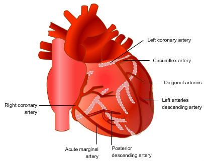
The major vessels of the coronary circulation are:
The left and right coronary arteries originate at the base of the aorta. The major coronary vessels lie on the epicardial surface of the heart and function as low resistance, distribution vessels. These epicardial arteries branch into smaller arteries that dive into the myocardium and become the micro vascular resistance vessels that regulate coronary blood flow. Arterioles give rise to a dense capillary network so that each cardiac myocyte has several capillaries running parallel to the muscle fiber. The high capillary-to-fiber density ensures short diffusion distances to maximize oxygen transport into the cells and removal of metabolic waste products from the cells (e.g., CO 2 , H + ).
